

Big plunging ranula which was diagned as lipoma or lymphangioma
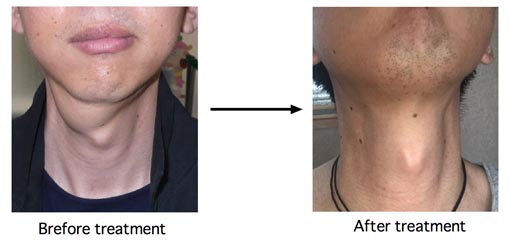
Young man. He first noticed right submandibuler swelling 2
years ago, Then he went to a hospital. ENT doctor told him that he had
llipoma and needed extirpation operation. He did not want to have
surgery, so he hesitated to be treated. Several months after, his right submandibuler
swelling was spontaneously decreased. But, 3 months ago, his right
submandibuler swelling became increased again. He went to another doctor and
had MRI study. Plastic surgeon told him thst he had lymphangioma and
needed very difficult extirpation operation.
He found my
website and came to me from very far place.
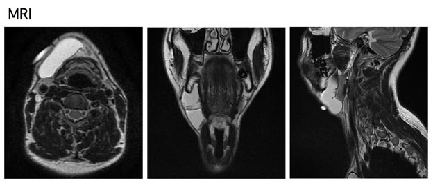
These are his MRI images. White lesion means
fluid
collection. I think this is
typical ranula MRI. And his ranula exists in front of submandubuler gland. This means OK-432 therapy is easy.
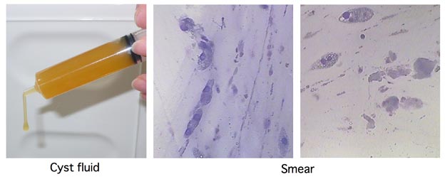
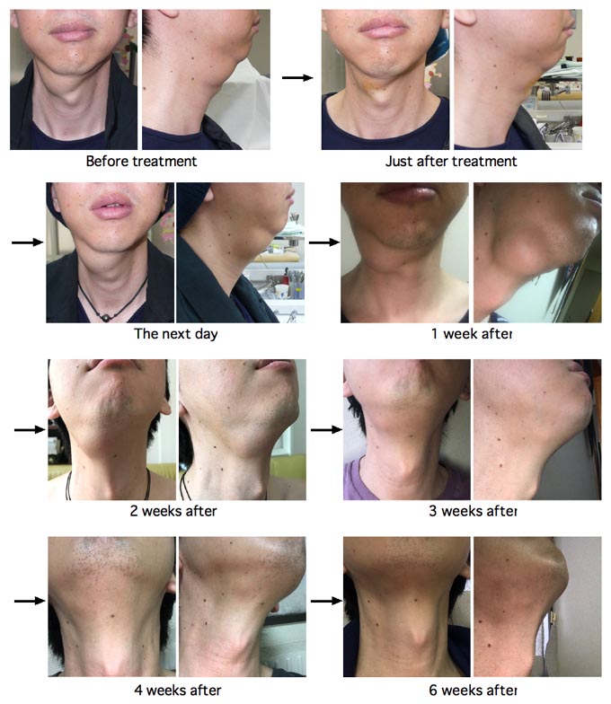
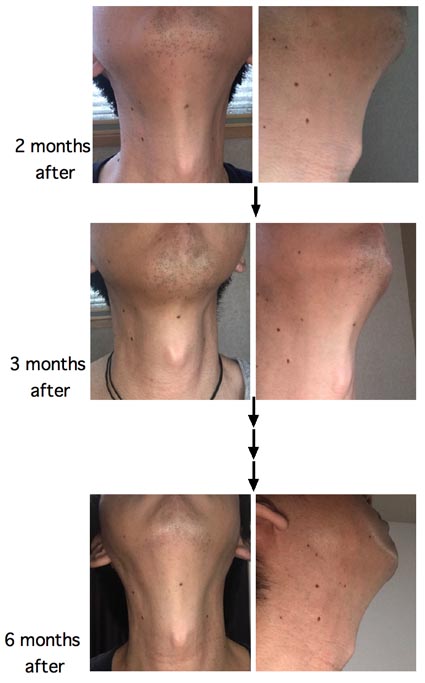
This is the course of his treatment. 12 ml
of cyst fluid was removed and then 2 ml of OK-432 susupensioin was
injected. So, his submandibular swelling was a little reduced at just
after treatment. But at the
next day, his ranula
was swollen. This means good response. 1 week after treatment, swelling
was maximum. After 2 weeks, his
ranula began to shrink. 6 weeks after treatment,
there was no swelling.
Please Click here to go to English Page with frame (If you can't see frame, click it)