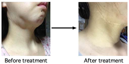

Ranula that was appeared after
removal of salivary stones and turned to be a plunging ranula after
several treatmens including removal of sublingual gland.

She had surgery on her neck when she was infant. The wound (scar) seen in
these pictures is due to this surgery. So, this wound has no relationship to
ranula.
She felt her right submandibuler swelling and tongue pain 2 years ago. There were salivary stones.
So, she had removal operation of salivary stones from mouth. After this
surgery, she had oral floor ranula. Her oral ranula repeated swellings
and raptures. So, she had removal operation of sublingual gland from mouth. But,
her ranula was not improved. So, she had drainage tube insertion
operation. But, after removing drainage tube, her ranula turned to be
a plunging ranula. So, her doctor recommended her to remove submandibular
gland. She worried that she would never improve and mailed me for asking
treatment of ranula.

Left is her MRI. White lesion means fluid
collection. The shape of white lesion is typical to ranula. Her ranula
seemed to be multi cystic. Right is
aspirated cyst fluid. Yellowish mucous fluid. This is typical for
ranula. After aspiration of 5 ml cyst fluid, 1 KE of OK-432 suspended
with 2 ml of saline was
injected.

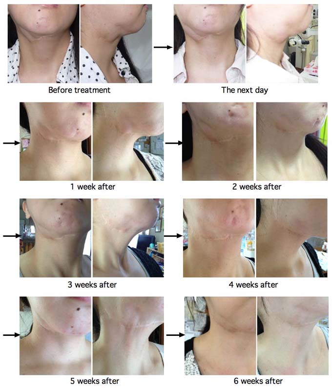
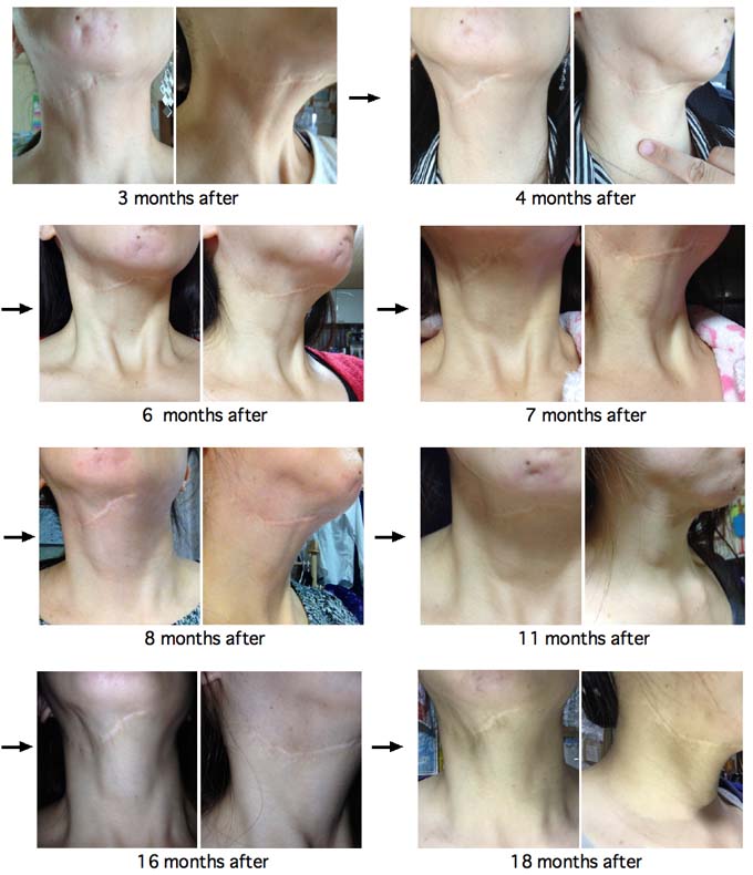
This is the course of her treatment. The
next day, her ranula
was
not swollen and the color of local skin did not become red. And fever
up was not observed. This dose not mean good
response. One
week after, her ranula became slightly hard, then gradually
became small. But, after 5 weeks, soft swelling appeared again.

Please Click here to go to English Page with frame (If you can't see frame, click it)