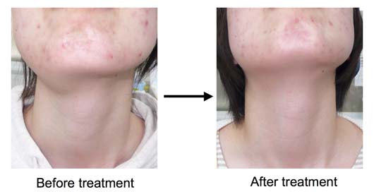


This patient noticed her right submandibular swelling from 2 years
ago. She did not go to doctor because she felt no pain. Two years after
she became particlar about appearence, then she went to an ENT doctor.
He thought that she must be plunging ranula then sent me.

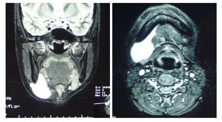
This is her MRI. White lesion means fluid collection. The shape of white lesion is typical for ranula.

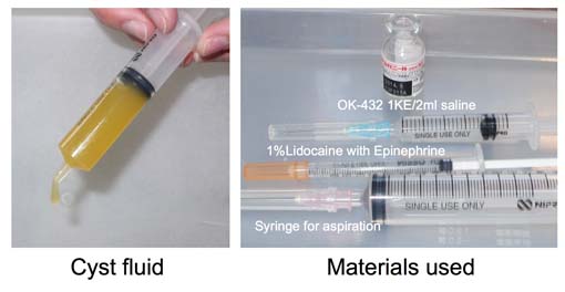
Left is her cyst fluid. Typical fluid of ranula.
Right is materials used. From upper side, vial of OK-432 1KE, OK-432
1KE susupended with 2ml of saline, 1% Lidocaine with epinephrine
(local anesthesia), and a 10ml syringe with 18G needle for aspiration.
I aspirated 10ml of cyst fluid and then injected all solution,
2ml, of OK-432.

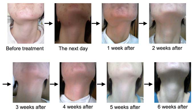
This is a course of treatment. The next day, her ranula was swollen and the color of local skin became red. One week after, her ranula was more hardly swollen. This means very good response. 2 weeks after treatment, her ranula began to shrink. 6 weeks after treatment, there was no swelling.
Please Click here to go to English Page with frame (If you can't see frame, click it)