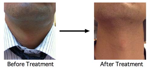


At first, he had ranula at his left oral floor. He went to the
hospital near his living place. He was only follwed up. After that, his
raula spontaneously changed to plunging type. Then, he was sent to
University Hospital. But, he was only followed up. He found my website
and came to me from Kanto district Japan. I treated him on one day trip.

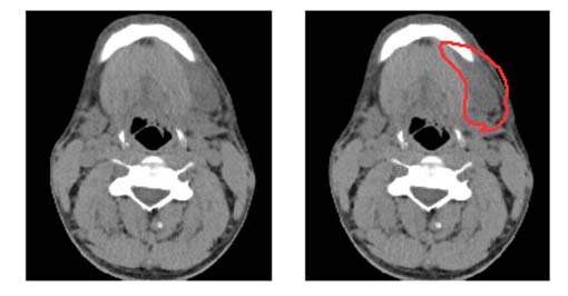
He had had only X-ray CT examination. This slice is most eminent part. The area that was enclosed by orange line is ranula. CT image of ranula is not so clear. I think T2 insisted image of MRI is the best for ranula imaging. From this CT, I could know that his ranula existed just under submandibular skin, this means that it is easy to treat.

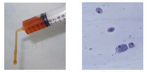
This is his cyst fluid. Slight red color means containg small amout of blood. Right is smear of cyst fluid 400X. These large cells are usually recognized in cyst fluid of ranula.

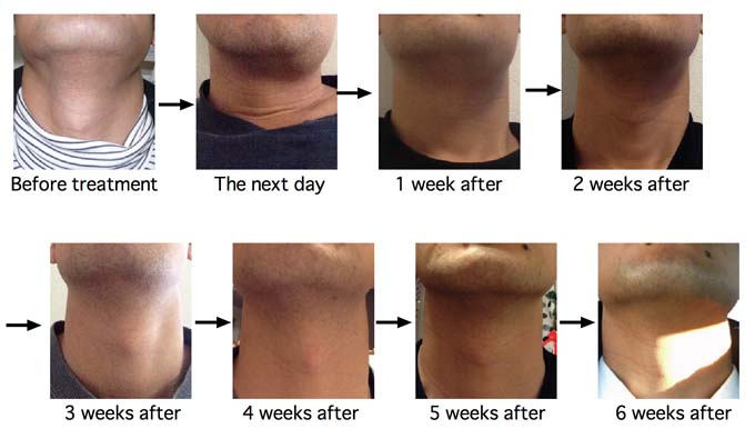
The next day after OK-432 injection, his
submandibular lesion was swollen and reddened. 1 week after, his left
submandibular swelling became hard. Then began to shriunk. 4 weeks
after treatment, swelling was disappeared.