

Ranula occured as a complication of sialoendoscopy
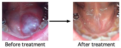
Adult woman. She noticed her left submandibular swelling 8 months before her first come to my clinic. She was suspected to be sialolithiasis. So, she had sialoendoscopy at an university hospital. The doctor could not find out her saliva exit, so sialoendoscopy was done under incision of Wharton's duct. But, there was no salivary stone. Her submandibular swelling gradually disappeared. But, one month after, ranula occured at her left oral floor. After that, she had two marspialization operations. But, her ranula became bigger and bigger.
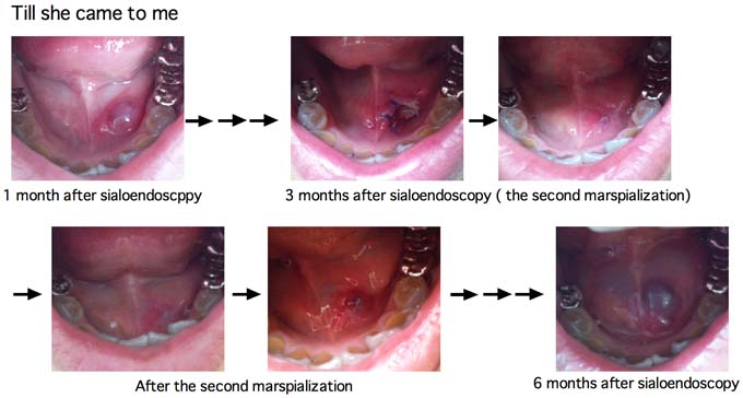
Upper photoes are attatched to her first consulting mail. Upper left
is the photo that ranula was appeared 1 month after sialoendoscopy. The
next four photoes are the second marspialization operation and after.
The last photo is 6 months after sialoendoscopy.
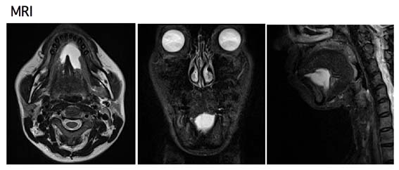
These are her MRI. White lesion is ranula. Ranula was big, but ranula was only localized at oral floor.
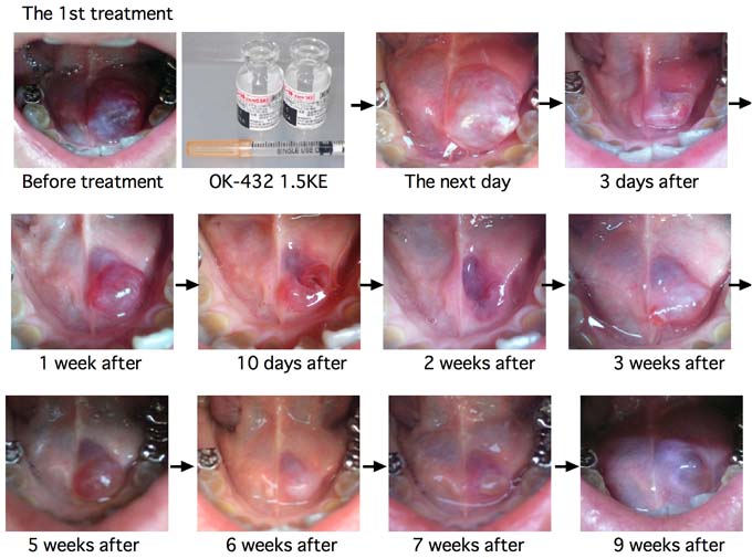
This is the 1st OK-432 treatment. 1,5 KE of OK-432 susupended with 0.3ml saline was injected both side of ranula. The next day, ranula was swollen and reddened. This means very good response, But, 3 days ater, her ranula was raptured. Then, her ranula repeated swellins and raptures. 7 weeks after, her ranula returned to be same size as that of before treatment.
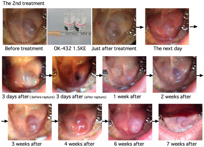
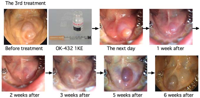
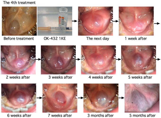

6 months after the 4th treatment, I got a
mail from her that oral floor swelling was not seen from several days
ago. After that, there was no recurrence for 4 months.
This course may be spontaneous healing. But, such a case that ranula
was disappeared after long duration from OK-432 injection was also
reported. In this HP. plunging raula 19, plunging ranula 29, combined ranula 2, and combined ranula 5 took the same course. This therapy needs patience to both patient and doctor.
Please Click here to go to
English Page with frame (
If you can't see frame, click it)